Elements of Visual Perception
The Human Eye.
The retina, pupil, iris, cornea, lens, and optic nerve are just a few of the approximately 40 distinct subsystems that make up the human eye, which is incredibly intricate and interconnected. For example, the retina contains about 137 million unique cells that react to light and communicate with the brain. These rod-shaped cells, which number about 130 million, are responsible for vision in black and white. We can see the colour thanks to the remaining seven million, which are cone-shaped. Light impressions are captured by the retinal cells, converted to electric pulses, and transmitted to the brain via the optic nerve. We can see "pictures" of our world because of a special part of the brain called the visual cortex, which interprets pulses to color, contrast, depth, etc. Amazingly, the visual cortex, optic nerve, and eye are all separate and distinct subsystems. Nevertheless, they can collectively receive, transmit, and decode up to 1.5 million pulse messages every millisecond.
The diameter of the eye is approx. 20 mm
There are 3 membranes enclosing the eye.
-
-
Cornea & sclera
-
Choroids
-
Retina
-
The Choroids.
The choroid contains blood vessels for eye nutrition and is heavily pigmented to reduce extraneous light entrance and backscatter. It is divided into the ciliary body and the iris diaphragm, which controls the amount of light that enters the pupil (2 mm - 8 mm).
The Lens:
The lens is made up of fibrous cells and is suspended by fibers that attach it to the ciliary body. It is slightly yellow and absorbs approximately 8% of the visible light spectrum.
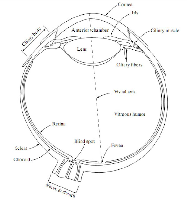
Figure. Cross section of the human eye.
The Retina :
The retina lines the entire posterior portion. The Discrete light receptors are distributed over the surface of the retina. There is two types of light receptor cones (6-7 million per eye) and rods (75-150 million per eye).
Cones: Cones are located in the fovea and are sensitive to color. Each cone is connected to its own nerve end and the cone vision is called photopic (or bright-light vision).
Rods: Rods are giving a general, overall picture of the field of view and are not involved in color vision. Several rods are connected to a single nerve and are sensitive to low levels of illumination (scotopic or dim-light vision).
Image Formation in the Eye
The eye lens (if compared to an optical lens) is flexible. It gets controlled by the fibers of the ciliary body and to focus on distant objects it gets flattered (and vice versa). The Distance between the center of the lens and the retina is called focal length it Varies from 17 mm to 14 mm.
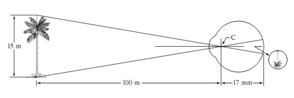
Figure: The eye looking at a palm tree Point C is the optical center of the lens
Calculation of retinal image of an object
The observer is looking at a 15 m high palm from a distance of 100 m. If h is the height in mm of that object in the retinal image, the (15*1000)/(100*1000)=h/17 or h=2.55mm. The retinal image is reflected primarily in the area of the fovea. Perception then takes place by the relative excitation of light receptors, which transform radiant energy into electrical impulses that are ultimately decoded by the brain.
Free online Tutorial, Free Online Notes, Digital Image Processing, DIP, Computer Vision, Analysis, Fundamental Steps, Image acquisition, Image Enhancement, Image restoration, Segmentation, Recognition, Components of an Image Processing System Elements of Visual Perception, Image Formation in the Eye, Dilation and Erosion, Opening and Closing, Noise, Noise, Pattern Recognition, Compression, Image compression, Lossy compression, Lossless compression
Digital Image Processing
DIP notes
DIP University Notes VTU AKTU
Digital Image Processing pdf
Digital Image Processing pdf notes
Digital Image Processing short notes
Digital Image Processing question paper
Digital Image Processing projects
Digital Image Processing projects using MATLAB
DIP Subject Notes PDF
Digital Image Processing Lecture Notes PDF